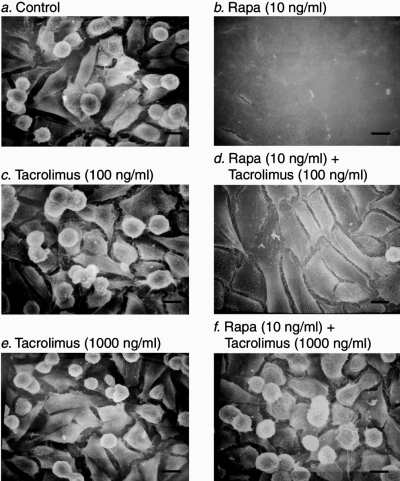
Fig. 2.
Conversion of renal cancer cells from an invasive phenotype to a non-invasive phenotype by rapamycin. Scanning electron microscope photographs of murine renal adenocarcinoma cells grown on glass cover slips and incubated for 72 h with: nothing (control cells, Panel A); 10 ng/ml rapamycin (Panel B); 100 ng/ml tacrolimus (Panel C); 10 ng/ml rapamycin and 100 ng/ml tacrolimus (Panel D); 1000 ng/ml tacrolimus (Panel E); 10 ng/ml rapamycin and 1000 ng/ml tacrolimus (Panel F). Note that rapamycin treatment induces phenotypic transition of renal cancer cells from spindle shaped cells with exploratory pseudopodia to cuboidal shaped cells that form cell-to-cell adhesion and display a cobblestone appearance, and that tacrolimus prevents the rapamycin-induced morphological alterations. Scale bars, 10 μm. (Reprinted from Luan et al (9) with permission).

