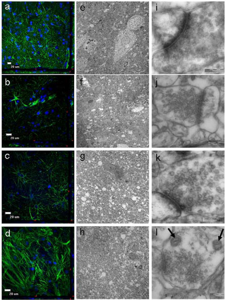
Figure 1.

Evaluation of normal adult human cortex obtained from a 46 year old female using electron microscopy (EM) and immuohistochemistry (IHC). a,e,i, 0 days in culture. b,f,i, 3 days in culture. c,g,k, 7 days in culture. d,h,l, 11 days in culture. The explants retained their cytoarchitecture for at least 11 days while in culture as seen by both EM and IHC. With the use of IHC analysis, the tissue cytoarchitecture remained intact through 11 days of culturing. This was evident by the numerous astrocytes and intact astrocytic processes. (a-d; GFAP-green, DAPI-blue). With the use of EM analysis, glial bodies appeared to persist through 11 days of culturing, and large vacuoles were notably absent, signifying surviving cells (e-h). At higher magnification, the cell membranes are maintained without distortion and the synaptic contacts are highly preserved (i-l). Moreover, both the presynaptic and postsynaptic vesicles can be clearly seen, as well as mitochondria (l: arrows) without altered morphology (e-h).
