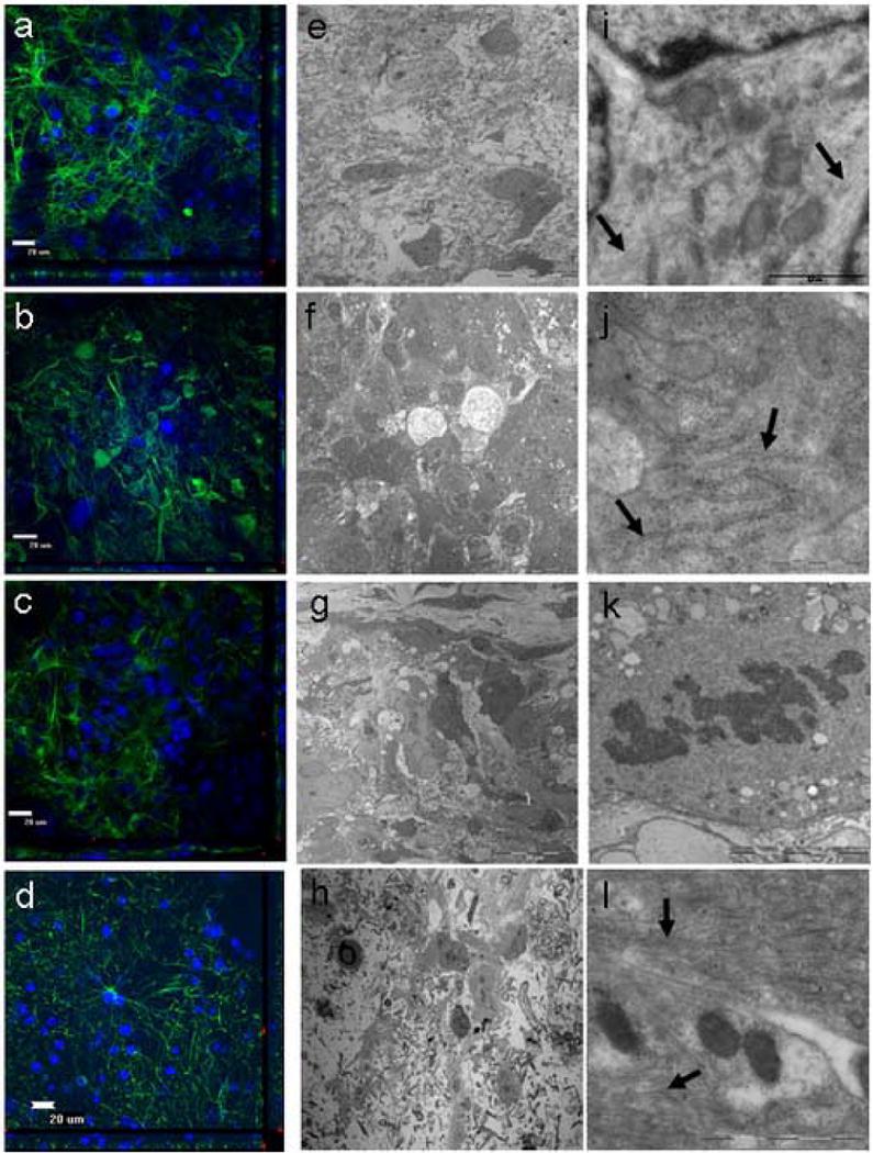
Figure 3.

Evaluation of human glioblastoma multiforme obtained from a 55 year old male using electron microscopy (EM) and immuohistochemistry (IHC). a,e,i, 0 days in culture. b,f,j, 4 days in culture. c,g,k, 6 days in culture. d,h,l, 10 days in culture. Unlike the normal cortex, the tumor cytoarchitecture appeared less organized and thus more characteristic of tumors (a-d; GFAP-green, DAPI-blue). The tumor explants retained their cytoarchitecture for at least 10 days while in culture as seen by both EM and IHC. With the use of EM analysis, the organization of the tumor cells in culture did not show any significant differences compared to tumor cells observed in vivo (e-h). At higher magnification, the characteristic organelles within the tumor astrocytes were preserved. This included intermediate filaments (i, l: arrows) and rough endoplasmic reticulum (j: arrows). Mitotic bodies were also seen in the explants, supporting the notion that the tumor explants were still proliferating and were remarkably preserved (k) (e-h). With the use of IHC analysis, the tissue cytoarchitecture from the tumor explants remained intact through 10 days of culturing.
