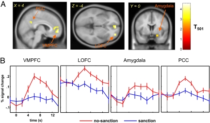
Fig. 3.
The trustee's brain regions showing greater activation in the no-sanction condition than in the sanction condition (P < .001, uncorrected; cluster size k > 5 voxels). (A) A random-effects GLM analysis reveals several brain regions significantly more activated by the revelation of no sanction. These regions include the VMPFC (peak activation MNI coordinate [4 56 −4]), right amygdala (peak activation MNI coordinate [24 0 −20]), right LOFC (peak activation MNI coordinate [32 52 −4]), and PCC (peak activation MNI coordinate [4 −24 36]). (B) Mean event-related time courses of the 4 brain regions. The dashed line indicates the time onset; error bars are SEM. Bold signals in the VMPFC, LOFC, amygdala, and PCC are all significantly greater when the trustee is in the no-sanction condition (red traces) than when she is in the sanction condition (blue traces).

