
FIG. 1.
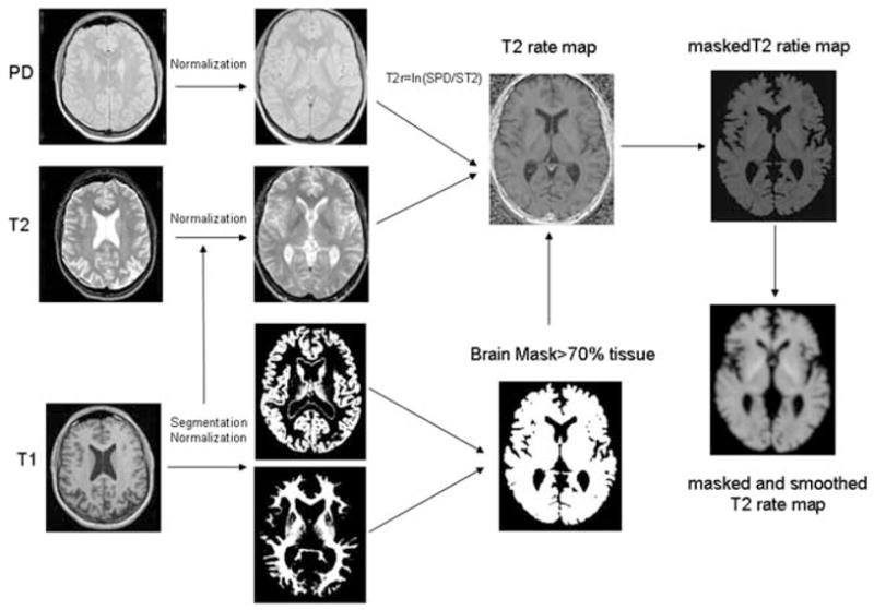
Postprocessing steps (see also Methods). The T1 was coregistered to T2/PD and the resulting image was used for segmentation with SPM2. The gray matter map was then normalized to the gray matter prior and the deformation parameters were applied to the coregistered T1, T2, and PD. The normalized T2 and PD were used to create T2 relaxation rate maps, which were masked with a tissue mask to reduce partial volume effects. For the analysis in SPM, the masked T2 relaxation maps were smoothed with a Gaussian kernel of 4 mm FWH.