
FIG. 2.
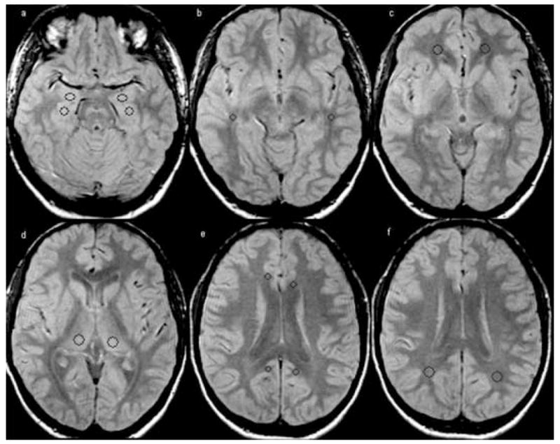
Placement of the regions of interest (ROI) in the hippocampus (size: 44 pixels, Talairach coordinates: −24, −14, −17 and 27,−14,−17) and, amygdala (size: 44 pixels, Talairach coordinates: −23,−4,−17 and 23,−4,−17) (a), temporal stem (size: 24 pixels, Talairach coordinates: −44,−30,−7 and 46,−30,−7) (b), orbitofrontal white matter (size: 61 pixels, Talairach coordinates: −21,42,−7 and 21,42,−7) (c), thalamus (size: 68 pixels, Talairach coordinates: −14,−23,8 and 18,−23,8) (d), posterior (size: 24 pixels, Talairach coordinates: −11,−46,23 and −7,−48,23) and anterior cingulate (size: 24 pixels, Talairach coordinates −10,29,19 and 10,25,19) (e) and parietal white matter (size: 68 pixels, Talairach coordinates: −28,−50,25 and 31,−50,25) (f).