Figure 3.
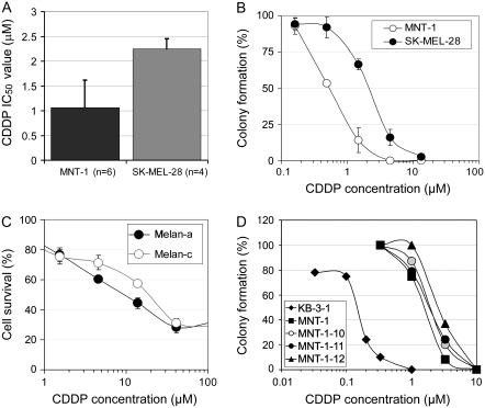
Melanosome stages, melanin content, and cisplatin (CDDP) sensitivity. A) CDDP resistance of SK-MEL-28 (containing stage I and II melanosomes) and of MNT-1 cells (enriched with stage III and IV melanosomes). IC50 values were determined from multiple independent experiments as indicated. Columns represent mean IC50 values and error bars 95% confidence intervals (CIs). B) Clonogenic assays of CDDP sensitivity in SK-MEL-28 and in MNT-1 cells maintained in high-density culture conditions. Approximately 1000 highly pigmented cells were seeded in 60-mm cell culture dishes. The cells were treated on the 3rd day (to ensure proper plating efficiency and vitality of the cells) with CDDP for 3 days. We counted cells in all dishes on day 12 after removal of the drug-containing medium in this experiment. Representatives of triplicate dishes of each treatment are shown in Supplementary Figure 3, A (available online). Cytotoxic dose–response curves were plotted, with each point corresponding to the mean value and error bars indicating 95% CIs. One of two similar experiments is shown. C) Drug sensitivity in immortal mouse melanocytes (melan-a, wild type) and their hypopigmented mutant (melan-c, albino). Approximately 5000 cells per well were seeded in 96-well plates. The cells were treated on the 3rd day (to ensure proper plating efficiency and vitality) with CDDP for 72–96 hours. Cytotoxicity was measured using the 3-(4,5-dimethylthiazol-2-yl)-2,5-diphenyl tetrazolium assay as previously described (28); each point corresponds to the mean value of quadruplicate determinations in each independent experiment and error bars indicate 95% CIs. One of two similar experiments is shown. D) Clonogenic assays of CDDP sensitivity in MNT-1 subclones. KB-3-1 cells (derived from the HeLa cervical adenocarcinoma cell line) were used as a nonmelanoma control. Approximately 300 cells were seeded in 60-mm cell culture dishes. The cells were treated with CDDP for 10 days. Cytotoxic dose–response curves were determined as described in (B). One of two similar experiments is shown. A comprehensive analysis of cytotoxic drug sensitivity and its association with melanin content is provided in Supplementary Table 1 (available online).

