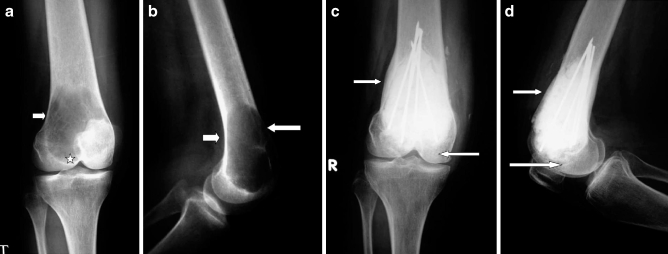
Fig. 6.
a, b Preoperative X-rays of a case of GCT of the distal femur (case 2) showing cortical thinning (arrow) and subchondral extension (star). c, d Follow-up X-rays showing internal fixation with intramedullary wires with 40 month post-reconstruction showing cortical and subchondral thickening (arrows)

