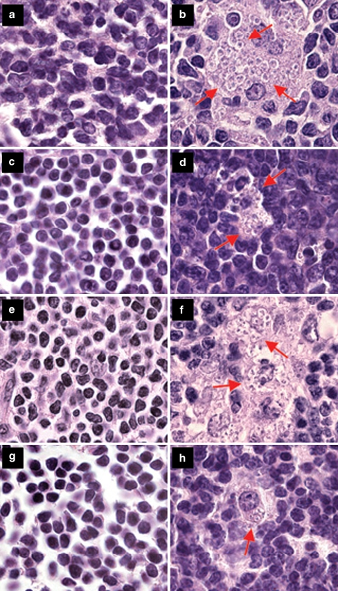
Fig. 1.
Parasites in hematoxylin–eosin-stained inguinal lymph node sections of female and male mice. a Uninfected BALB/c male, bL. major infected BALB/c male, c uninfected BALB/c female, dL. major infected BALB/c female, e uninfected CcS-11 male, fL. major infected CcS-11 male, g uninfected CcS-11 female, hL. major infected CcS-11 female. Arrows show groups of Leishmania parasites (with a dark nucleus, smaller kinetoplast, and light cytoplasm) among normal lymph node cells (b, d, f) and parasites inside a macrophage (h)

