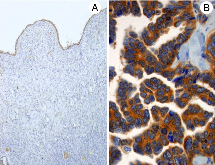
Fig. 2.

Staining for PRK1 was seen in normal ovarian surface epithelium (A) and ovarian serous carcinomas (B). Positivity for the serous carcinomas was usually strong, while that for the normal ovarian epithelium varied from negative/weak to moderate.
