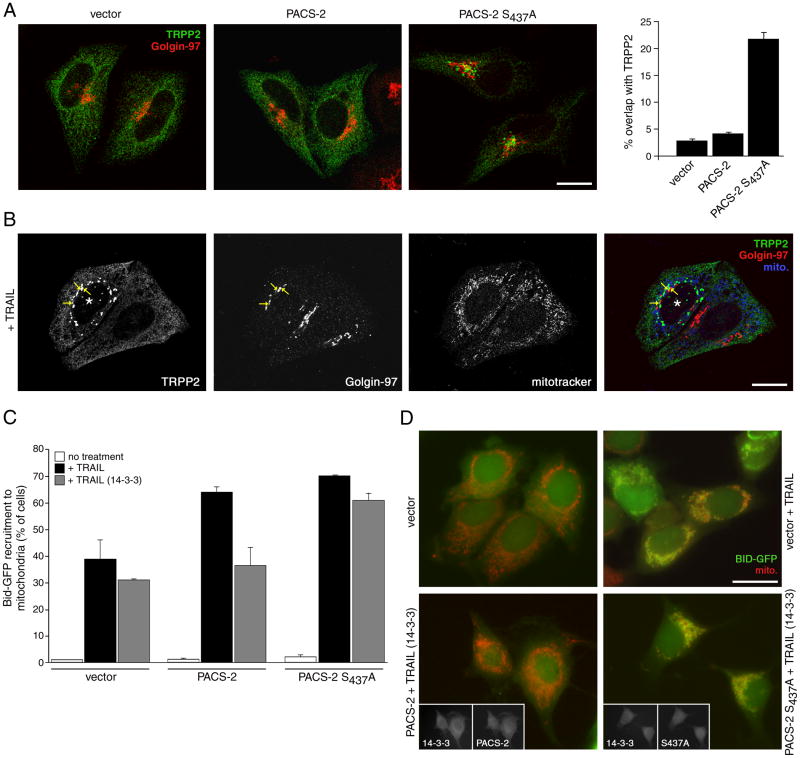
Figure 7. PACS-2 Ser437 and 14-3-3 integrate membrane traffic with apoptotic Bid action.
(A) HeLa cells were nucleofected (Amaxa) with myc-TRPP2 together with either empty vector or HA-tagged PACS-2 or PACS-2S437A. After 36 hr, the cells were fixed and processed for confocal microscopy using anti-myc (mAb 9E10, green) and anti-Golgin-97 (red) and visualized using Alexa-conjugated secondary antibodies. Right: Morphometric analysis. Error bars represent mean +/- SEM. Scale bar, 20 μm. (B) HeLa cells expressing myc-TRPP2 were treated with 20 ng/ml TRAIL for 4 hr and processed for confocal microscopy. Mitotracker (pseudocolored blue), anti-myc (green) and anti-Golgin-97 (red) are shown. *, apoptotic cell showing puncate myc-TRPP2 and collapsed mitochondria. Arrows, overlap of myc-TRPP2 and Golgin-97. Scale bar, 20 μm. (C) MCF-7:Bid-GFP cells were nucleofected (Amaxa) with pcDNA3.1 (vector) or plasmids expressing HA-tagged PACS-2 or PACS-2S437A alone or with 14-3-3ζ-myc. The cells were then treated with vehicle or 20 ng/ml TRAIL for 4 hr. Left: The percent of cells containing Bid-GFP translocated to mitochondria (mitotracker, red) in random fields (300 cells minimum) were quantified. Data are represented as mean +/- SD. Right: Immunofluorescence of MCF-7:Bid-GFP expressing the indicated proteins and treated or not with TRAIL. Insets, co-expression of 14-3-3ζ-myc with HA-tagged PACS-2 or PACS-2S437A. Scale bar, 20 μm.

