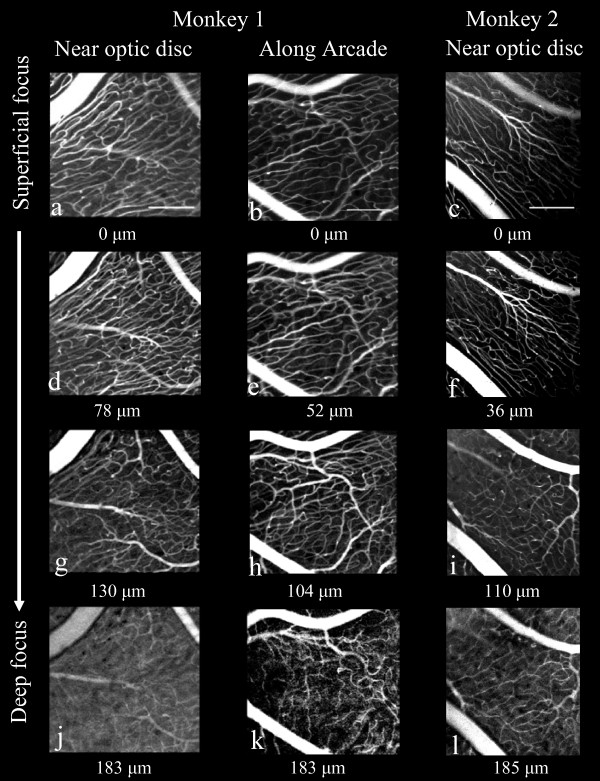
Figure 3.
Through-focus series of in-vivo adaptive optics images of RPCs from two retinal locations in monkey 1 and one retinal location in monkey 2. One location in both monkeys is near the optic disc; monkey 1 (a, d, g, j) and monkey 2 (c, f, i, l) and the second is 5 mm temporal to the optic disc of monkey 1(b, e, h, k) (see Figure 1 for locations in monkey 1). At a focal plane deep within the NFL, a transition was found from RPC to to the typical, polygonal ganglion cell layer/retinal circulation (j, k, l). The focus depth set by the deformable mirror relative to the most superficial vasculature is displayed under each image. Scalebars indicate 200 μm.

