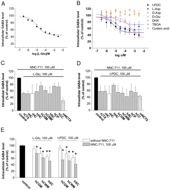
Figure 1. Cytosolic GABA level as determined by [3H]GABA uptake in the presence of 100 µM NNC-711.
(A) Inhibition of [3H]GABA uptake following 10 min preincubation with Glu in rat cerebrocortical NPMV fraction (n = 7). IC50 value for Glu: 11.0±0.1 µM. (B) Inhibition of [3H]GABA uptake following 10 min preincubation with different EAAT substrates and inhibitors in rat cerebrocortical NPMV fraction (n = 3–6). (C and D) Decreased intracellular GABA level following Glu (C) or t-PDC (D) application in rat and human NPMVs (gray), from acute rat hippocampal slice (empty bar, n = 7) and from neuronal culture from mouse neocortex (striped bar, n = 4). Abbreviations used for brain regions: rat cerebrocortical neocortex (rNCTX, n = 6), rat hippocampus (rHC, n = 3), human hippocampus (hHC, n = 6), human cortical gray matter (hCGM, n = 6), human cortical white matter (hCWM, n = 6), human spinal cord white matter (hSC, n = 6), human choroid plexus (hCP, n = 6), human corpus callosum (hCC, n = 6). All drug applications significantly differ from the control (P<0.01). (E) Intracellular [3H]GABA level in the absence and presence of 100 µM NNC-711 during application of Glu (100 µM) or t-PDC (100 µM) in NPMV fractions from human cortical gray matter (hCGM, n = 6), human cortical white matter (hCWM, n = 6) and human spinal cord white matter (hSC, n = 6). Asterisks: P<0.05.

