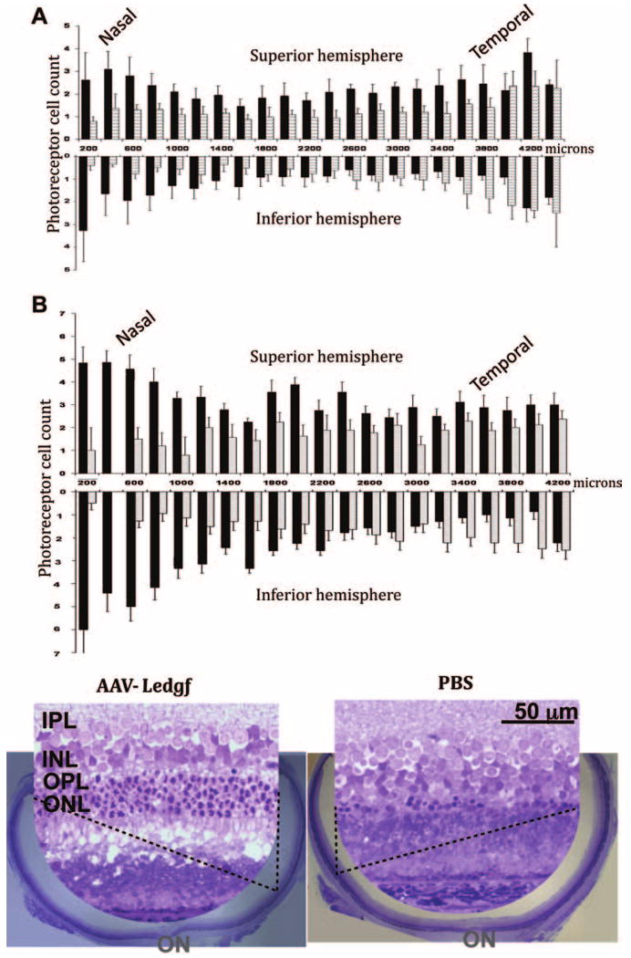
Figure 4.

Morphologic evidence of photoreceptor protection. (A) Average photoreceptor cell counts in the superior and inferior halves of radial retinal sections taken every 200 μm perpendicular to the nasal–temporal plane. AAV-Ledgf–treated (n = 5, ■) and PBS treated (n = 4,  ) rats were injected at 3 weeks of age and evaluated 8 weeks later. Standard error bars are shown. (B) Photoreceptor cell count in a representative AAV-Ledgf–treated eye (■) from the group presented in (A) and its contralateral PBS–treated eye (
 ) across the retina, and the corresponding retinal morphology in sections through the optic nerve. Insets are from the superior hemisphere. OPL, outer plexiform layer; ON, optic nerve head.
