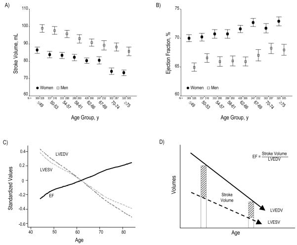
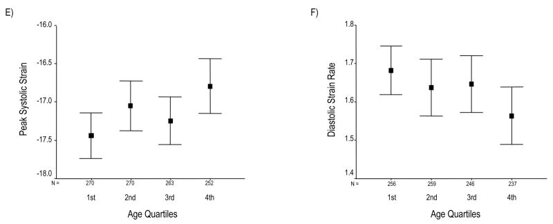
Figure 2.
SV declines across increasing age group (Panel A). Conversely, EF progressively rises (Panel B). Panel C summarizes the relation of age to these global functional indices by showing age-associated changes in standardized values for LVEDV, LVESV, and EF. Panel D depicts a schematic overview of changes in LV volumes and EF with increasing age. Although SV progressively falls, EF paradoxically increases; this appears due to the progressive decline in all LV volumes, particularly LVESV. Panel E depicts peak systolic strain (Ecc), reflecting the magnitude of myocardial systolic deformation, across increasing age quartiles. As age increases, midwall circumferential shortening becomes less negative, i.e. decreases since sysolic strain is conventionally negative during systole. Because diastolic strain rate is conventionally a positive value, reduction of its magnitude reflects slower circumferential lengthening during LV filling or progressively reduced myocardial relaxation with age (Panel F). Values in panels A, B, E, and F are presented as means (95% CI).


