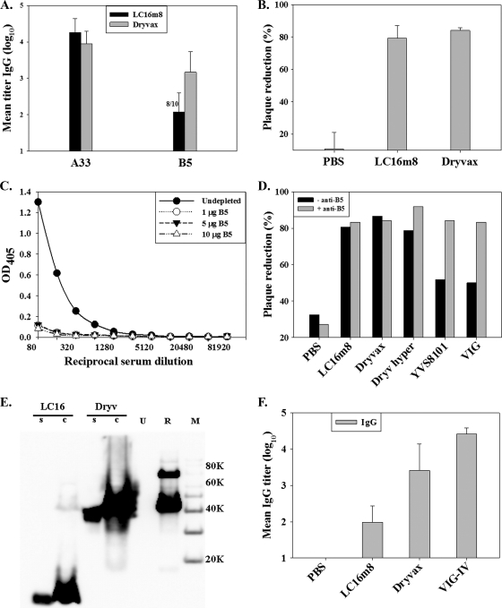
FIG. 2.
Antibody response to vaccinia virus EV proteins. Groups of five mice were vaccinated by tail scarification. (A) Serum samples were obtained from mice 6 weeks after vaccination and tested for A33-specific and B5-specific IgG. (B) Serum pools were diluted 1:50 and tested for neutralization of vaccinia virus EV by plaque reduction. (C) Depletion of B5-specific IgG from Dryvax hyperimmune serum was verified by ELISA. (D) Serum pools from mice inoculated with PBS, LC16m8, Dryvax, Dryvax hyperimmune serum (Dryv hyper), rabbit anti-vaccinia virus antibody (YVS8101), and human VIG were depleted (− anti-B5) or undepleted (+ anti-B5) of B5-specific IgG and tested for the neutralization of vaccinia virus EV. OD405, optical density at 405 nm. (E) Expression of the truncated B5 protein in LC16m8 (LC16)-infected or Dryvax (Dryv)-infected RK-13 cell supernatant (s) or cell lysate (c) was detected by Western blotting using a monoclonal antibody, NR-561 (1). Lane U represents uninfected-cell lysate, lane R represents affinity-purified recombinant B5, and lane M is the protein molecular weight marker. (F) Immune serum sample pools were tested by ELISA for levels of IgG to the truncated B5 using a recombinant truncated B5 protein as an antigen. Error bars represent standard deviations.

