Figure 3.
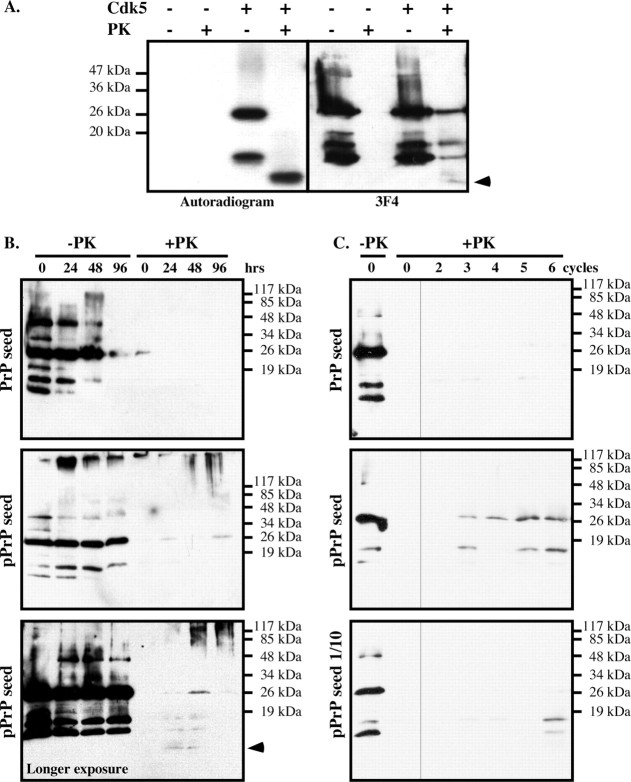
Cdk5-phosphorylated PrP induces the aggregation of nonphosphorylated PrP in vitro. A, Autoradiogram and Western blot analysis with 3F4 antibody of nonphosphorylated or Cdk5-phosphorylated PrP treated with 10 μg/ml PK for 1 h at 37°C. The arrow indicates the 10 kDa PrPRES fragment detected on the autoradiogram or Western blot. B, PrP Western blot of nonphosphorylated PrP seeded with kinase assays performed with (pPrP) or without Cdk5 and incubated for the indicated time without (−PK) or with (+PK) PK treatment. The bottom shows a longer exposure of another pPrP seeded experiment revealing the 10 kDa PrP fragment in +PK. C, PrP Western blot of nonphosphorylated PrP seeded with a 2 μl aliquot of the 96 h time point (0 cycle) in B and incubated 24 h (cycle 1). Subsequent cycles represent samples in which 2 μl at the end of the incubation period was added into fresh nonphosphorylated PrP and incubated 24 h. The bottom represents an original seed of 0.2 μl of the 96 h time point in B.

