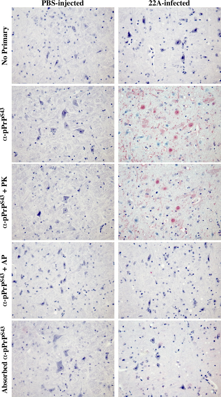
Figure 7.

Immunohistological staining of control PBS- and 22A scrapie-infected mice brains. Micrographs of control PBS-injected or 22A-infected mice brain tissue sections of the medulla untreated (no primary, anti-pPrPS43, adsorbed anti-pPrPS43), pretreated with PK (anti-pPrPS43 + PK), or pretreated with alkaline phosphatase (anti-pPrPS43 + AP) with anti-pPrPS43, no primary antiserum, or adsorbed anti-pPrPS43.
