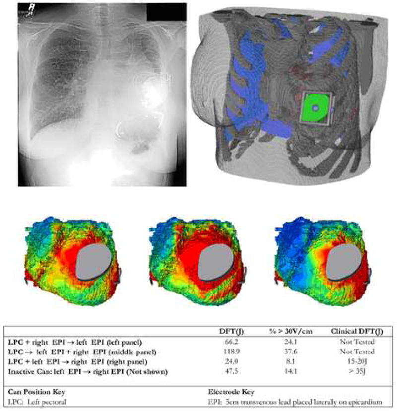
Figure 7. Patient specific modeling in patient with congenital heart disease.

Top: Post-implantation chest X-ray and corresponding finite element model showing can and epicardial coil electrode placement. Middle: Epicardial voltage gradient distribution for three alternative shock vectors tested using model. Bottom: Predicted and observed defibrillation energies.
