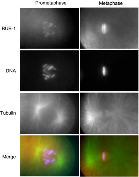
Figure 1. C. elegans has holo-kinetochores.
The conserved spindle assembly checkpoint protein BUB-1 kinase localizes to the diffuse C. elegans holo-kinetochore. Immunostaining of C. elegans embryonic cells at prometaphase (left column) and metaphase (right column) with anti–BUB-1 antibody and anti-tubulin antibody is shown. DNA is also visualized by staining with DAPI. The merged images of BUB-1 (red), DNA (blue), and tubulin (green) are shown in the bottom panel. In prometaphase, BUB-1 localizes along the entire length of each pair of sister chromatids. In metaphase, sister chromatids are highly compacted and congressed at the metaphase plate, where BUB-1 localizes the pole-ward face of each chromosome, thereby forming the classic two-threads shape. The anti-BUB-1 antibody for immunostaining was kindly provided by A. Desai.

