
Figure 4. Subcellular localization of MDF-1/CeMAD-1.
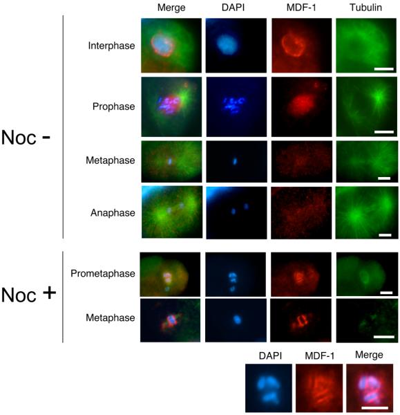
Fluorescence micrographs of early-stage wild-type embryos undergoing mitosis are shown. At this stage, MDF-1/CeMAD-1 localizes to kinetochore regions only in the presence of spindle damage. Wild-type embryos were dissected from adult hermaphrodites and either incubated for 15 min in 30 μg/ml nocodazole (Noc+) to induce kinetochore localization of MDF-1 or not incubated (Noc−). The embryos were then fixed and stained with DAPI (blue), anti–MDF-1 antibody (red), and anti-tubulin antibody (green). Images of chromosomes in the Noc+ cells in prometaphase are shown at the higher magnification in the bottom row. Scale bars: 10 μm.