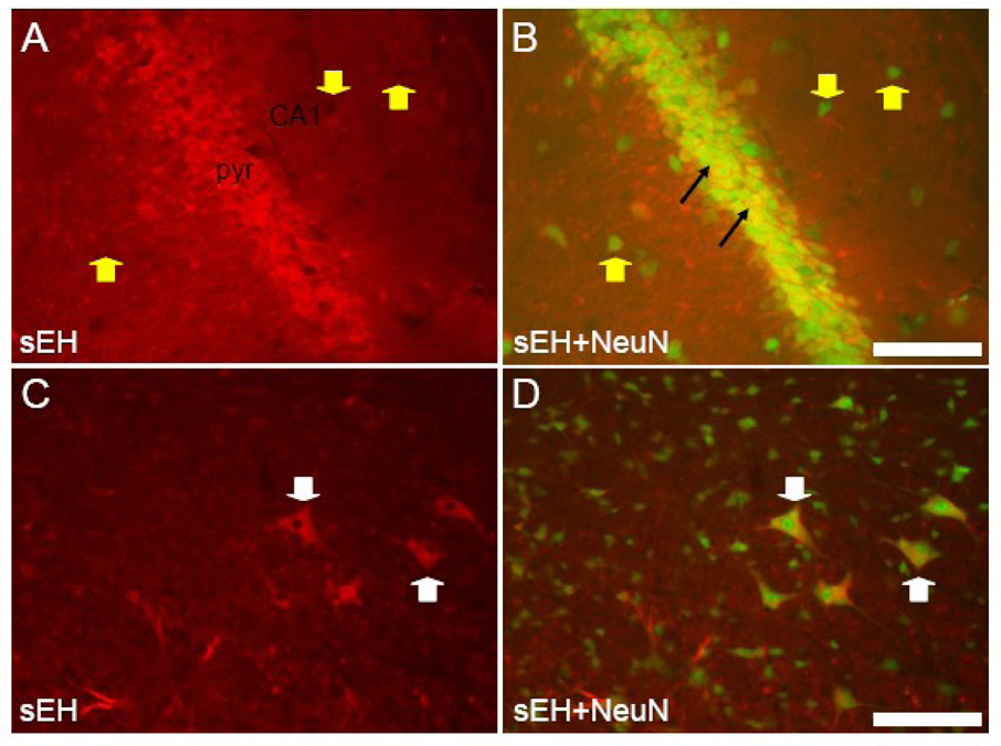
Figure 11. sEH is expressed in neurons of hippocampus and medulla in Syn-FLAG-sEH transgenic mice.
A and B. Coronal section through the CA1 region of a transgenic mouse hippocampus labeled with anti-sEH (A) and both anti-sEH anti-NeuN (B). Note the dramatic increase in sEH expression in pyramidal neurons (black arrows) compared to non-transgenic animals (e.g. Figure 4) though some neurons outside the pyramidal cell layer (yellow arrowheads) do not express sEH. C and D. Coronal section through the ventral medulla from a transgenic animal. Note the expression of sEH is still restricted to gigantocellular neurons (white arrowheads) which are clearly labeled by both anti sEH and NeuN antibodies. Scale bars = 100 µm

