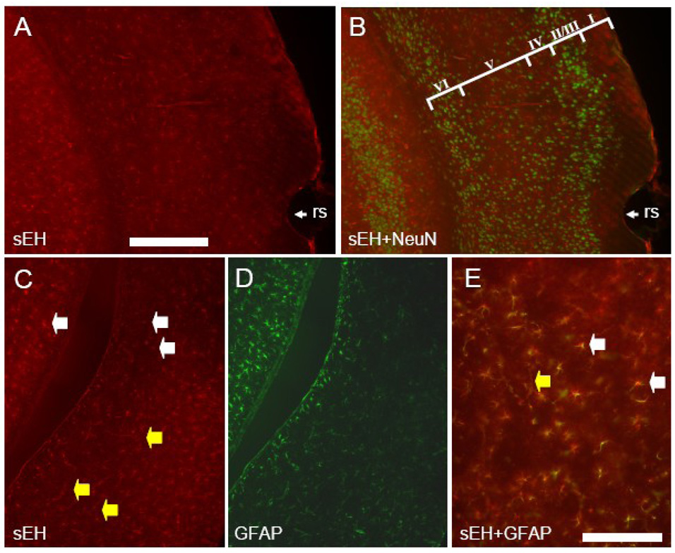
Figure 2. sEH is expressed in astrocytes and vascular cells in the lateral and ventral cerebral cortex.
Coronal section showing the lateral cerebral cortex dorsal to the rhinal sulcus (rs) labeled with anti-sEH (A) and anti-NeuN (B). Note no double labeled cells are present. Cortical layers are identified by roman numerals in panel B. Coronal section through the cerebral cortex ventral to the rhinal sulcus stained with anti-sEH (C) and GFAP (D). Higher magnification of merged C and D (E) shows extensive co-localization of sEH and GFAP (yellow cells). White arrowheads identify sEH positive astrocytes whereas yellow arrowheads identify sEH positive blood vessels. Scale bars: A–D = 500 µm; E = 100 µm

