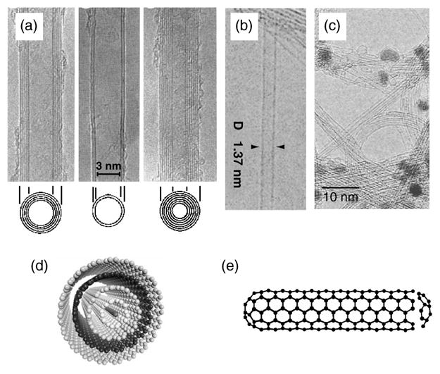
Fig. 1.

TEM micrographs of (a) MWNTs7 and (b) SWNTs8 (c) TEM micrograph showing bundles of SWNTs. The dark spots are catalyst particles used for nanotube growth. (d, e) Schematics of (d) MWNT and (e) SWNT. The TEM micrographs shown in (a) and (b) were kindly provided by Sumio Iijima, NEC, Japan and reproduced with the permission of Nature Publishing Group.
