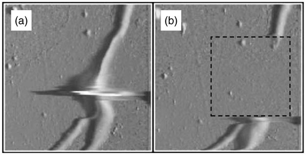
Fig. 12.

AFM images of neurites (~500 nm in diameter) containing a varicosity. (a) Deflection mode AFM image; imaging force, 12 nN. Artifact at neurite varicosity is due to delay in the feedback loop. The box in (b) indicates the area that was scanned at 72 nN after acquisition of (a) in order to remove a portion of the neurite. (b) Image of neurites after excision; imaging force, 12 nN.
