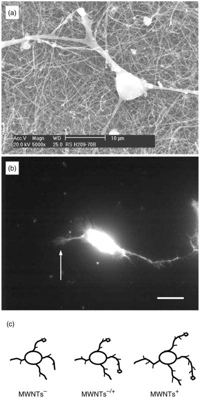
Fig. 14.

(A) SEM image of a neuron grown on as-prepared MWNTs. (b) Fluorescent image showing a live neuron on as-prepared MWNTs, which accumulated the vital stain, calcein. Arrow indicates a growth cone. Scale bar, 20 μm. (c) Drawing summarizing the effects of MWNT charges on growth cones, neurite outgrowth and branching. Modified from Ref. 106.
