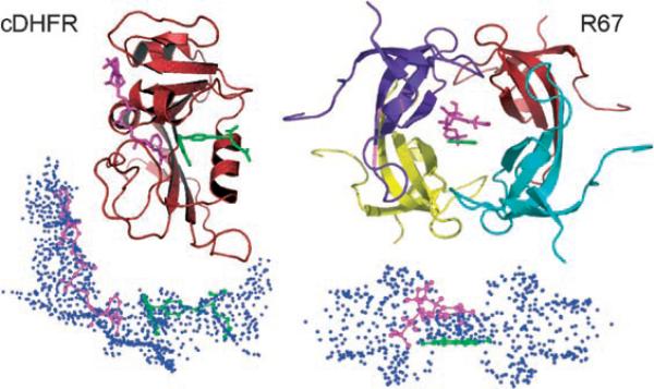
Figure 1.
Top: Structures of cDHFR (PDB ID R1X2; left) and R67-DHFR (PDB ID 1VIF; right). For R67-DHFR, the pteridine ring position (green) of the folate was defined in the crystal structure and the nicotinamide (magenta) was docked in place using DOCK. Bottom: Under each structure we present the reverse images for each active site.[18] In the reverse images, each sphere point describes a potential atom position for use by the docking algorithm. The sphere cluster for cDHFR is shown in approximately the same orientation as its structure. In contrast, the sphere cluster for R67-DHFR is shown sideways, after a 90° rotation along the y axis.

