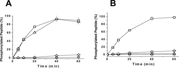
Figure 4.
Phosphorylation of the FXFP substrates by p38α kinase and JNK1. The designed substrates (1 μM) were incubated with the respective MAP kinase in the presence of ATP and Mg2+. Aliquots were removed at varying time points and amount of phosphorylated peptide was measured. A) Phosphorylation of the FXFP substrates by p38α kinase. Substrates with the most phosphorylation were ERKFXFP (triangles) and MEK1ERKFXFP (squares). Substrates with the longest time for phosphorylation were MEK1ERK (diamonds) and ERKSub (circles). B) Phosphorylation of the FXFP substrates by JNK1. Substrate with the most phosphorylation was MEK1ERKFXFP (squares). Substrates with the least phosphorylation was ERKFXFP (triangles), MEK1ERK (diamonds) and ERKSub (circles). Data points represent average of three measurements and error bars indicate their standard deviation.

