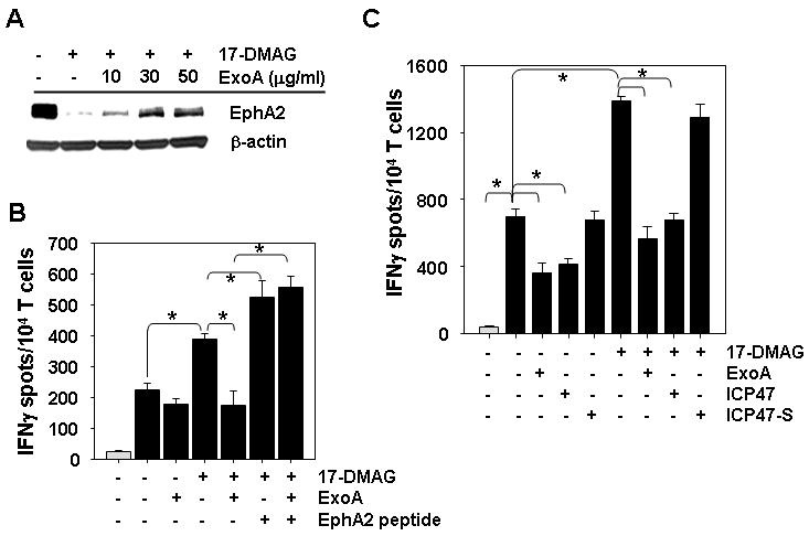
Figure 4. 17-DMAG treatment improves recognition of the SLR20.A2 RCC cell line by a low avidity, anti-EphA258-66 CD8+ T cell clone in a TAP- and retrotranslocation-dependent manner.

A. SLR20.A2 tumor cells were untreated or treated for 24h with 500 nM 17-DMAG +/- 10, 30 or 50 μg/ml ExoA, then analyzed for expression of EphA2 vs. control β-actin protein by western blotting. B. Untreated SLR20 cells (light gray bars) or SL20.A2 cells (black bars) pretreated as in panel A using 10 μg/ml ExoA were used as targets for recognition by anti-EphA258-66 CD8+ T cell clone 15/9 in IFN-γ ELISPOT assays. Drug-treated tumor cells pulsed with exogenous EphA258-66 peptide (10 μg/ml) to clone 15/9 was included as a positive control. C. SLR (light gray bars) and SLR20.A2 (black bars) tumor cells were untreated or treated with 500 nM 17-DMAG +/- 50 μg/ml ExoA, 10 μg/ml ICP471-35 peptide or 10 μg/ml ICP4735-1 scrambled peptide for 24h, prior to use as target cells for clone 15/9 T cell recognition in IFN-γ ELISPOT assays. All ELISPOT data are reported as the mean +/- SD of triplicate determinations. In all cases, data are representative of that obtained in 3 independent assays performed. * p < 0.05 for all indicated comparisons.
