
Figure 5. Thioflavine-stained Plaque Count.
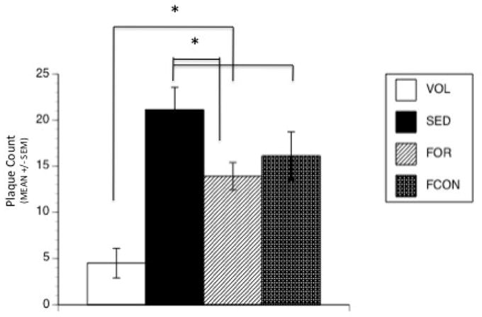
Quantative analysis of Thioflavine S stained plaques in the brains of Tg2576 mice at 9 months of age. Plaque number was significantly decreased in VOL group compared to all other groups. The FOR group had fewer plaques than the SED group. (* denotes p < 0.05).