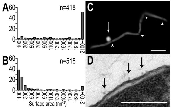
Figure 5. Quantitatively distinct septal- and cell body-derived MV populations.
(A) Septal-derived MVs, harvested from septate filamentous cells, are significantly larger than WT MVs (p=1.1×10-92), and (B) non septal-derived MVs, harvested from non-septate filamentous cells, are significantly smaller than WT MVs (p=1.3×10-49). (C) Large MVs are released at constricted septa (Bar = 5um), and (D) small MVs are cell body derived (Bar=200nm). Arrowheads highlight constricted septa and arrows highlight MV release; n = number of individual vesicles measured.

