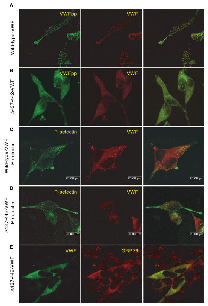
Fig. 2.

Intracellular localization of Δ437--442-von Willebrand factor (VWF). AtT-20 cells were transfected with wild-type (WT) VWF (A, C) or Δ437--442-VWF (B, D, E), or cotransfected with P-selectin (C, D). Cells were dual-stained for VWF propeptide (VWFpp) and VWF (A, B), P-selectin and VWF (C, D), or VWF and GRP78 (E). Intracellular localization was examined by confocal microscopy. The merged image is shown in the last column, with colocalization shown in yellow. Δ437--442-VWF does not form granules or colocalize with coexpressed P-selectin, but is instead localized to the endoplasmic reticulum.
