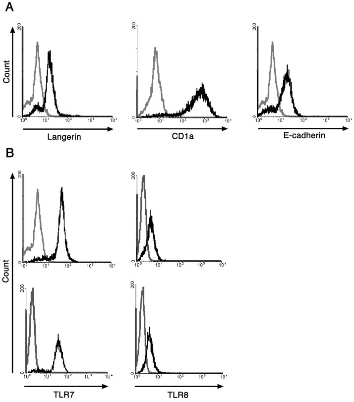
Figure 1. Characterization of monocyte-derived LC.
A. Monocyte-derived LC were stained with either anti-langerin, anti-CD1a, anti-E-cadherin (black histograms) or isotype matched negative controls (grey histograms). The cells were analyzed by flow cytometry. LC generated from monocytes express langerin, CD1a, and E-cadherin. B, Monocyte-derived LC were left untreated or exposed to HPV16 VLP and then permeabilized, fixed, and stained with either anti-TLR7, anti-TLR8 antibodies (black histograms) or isotype matched negative controls (grey histograms). The cells were analyzed by flow cytometry. Immature LC and LC exposed to HPV16 VLP express similar levels of TLR7 and TLR8. One representative experiment of three is shown.

