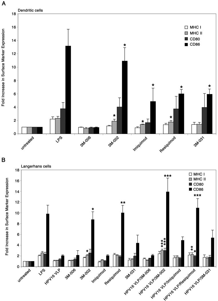
Figure 2. Differential expression of surface markers on DC and LC stimulated with imidazoquinolines.
A, DC were left untreated, treated with LPS, or treated with each of the imidazoquinolines. The cells were analyzed by flow cytometry for the expression of MHC class I and II molecules, CD80, and CD86. Surface markers are up-regulated when treated with 3M-002, imiquimod, resiquimod, and 3M-031. These data are represented by fold increase in surface marker expression, which are based on mean fluorescence intensity. The mean ± SEM of four separate experiments is presented (*P < .05). B, LC were left untreated, stimulated with LPS, exposed to HPV16 VLP, treated with each of the imidazoquinolines, or exposed to HPV16 VLP and subsequently treated with each of the imidazoquinolines. After the final incubation the cells were analyzed by flow cytometry for the expression of MHC class I and II molecules, CD80, and CD86. 3M-002 and resiquimod induced the up-regulation of surface markers on LC and LC exposed to HPV16 VLP. These data are represented by fold increase in surface marker expression, which are based on mean fluorescence intensity. The mean ± SEM of four separate experiments is presented (*P < .05, **P < .01, ***P< .001).

