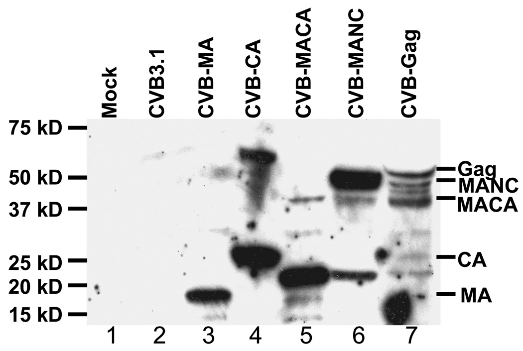
Figure 2. Expression of HIV-1 proteins by recombinant CVB3 vector.
Membrane probed with pooled serum from chronically HIV-1-infected patients. A549 cells were infected with lysates from cells transfected with each of the CVB3 constructs. Lane 1: Mock infection. Lane 2: CVB3.1. Lane 3: CVB-MA. Lane 4: CVB-CA. Lane 5: CVB-MACA. Lane 6: CVB-NC. Lane 7: CVB-Gag. Markers to the right indicate proper size of 3C-processed insertions of HIV-1 protein. Lanes 3 and 4 show MA and CA as expected size proteins after cleavage from the coxsackievirus polyprotein. Lane 5 indicates MACA expression of the expected 41 kD protein as well as a predominant 20 kD truncated protein. Lane 6 shows that the CVB-MANC construct expresses protein of the expected size (48 kD), but also indicates a less intense 20 kD truncated protein. Lane 7 shows the full length HIV-1 Gag protein and multiple truncation products.

