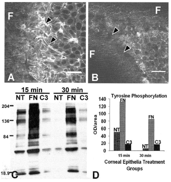
Figure 3.

Corneal epithelia treated with exoenzyme C3 (3 μg/mL) did not reorganize the ACM (B) compared with the control (A) and decreased tyrosine phosphorylation (C, D) compared with FN-stimulated epithelia. The total optical density from this sample immunoblot using the whole lane demonstrated that C3 decreased tyrosine phosphorylation 8- to 10-fold (D). All experiments and procedures were repeated at least three times with similar results. Single confocal optical sections through basal cells, as illustrated in the tissue schematic (Fig. 2), show the distribution of the F-actin in epithelia stimulated with FN in the control tissues (A). Areas that did not contain actin staining (dark) were optical sections through the supporting filter (F). The tissues were not flat; therefore, a single optical section may contain the filter (F) and basal cell cytoplasm. Scale bar, 20 μm.
