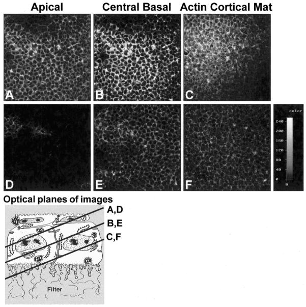
Figure 4.
Corneal epithelial cells were transfected with sense or antisense oligonucleotides directed toward Rho, immunolabeled with anti-Rho antibody, and analyzed with by CLSM, to determine intracellular distribution and transfection efficiency. The pinhole, voltage, and offset were the set at the same levels on both images. The intensity wedge indicates the range of detected light intensity from 0 (black) to 255 (white). Rho in control sense-transfected epithelia had a honeycomb distribution, similar to other membrane-associated proteins (A–C). The protein was less prominent in the periderm cells but was evenly distributed throughout the basal cells in a membrane-bound pattern (A–C). In contrast, epithelia transfected with antisense Rho had significantly less immunolabeled protein, indicating that the transfection decreased the amount of Rho protein evenly throughout the epithelium (D–F). The intensity wedge indicates the relative intensity of the pixels in each image. The schematic drawing indicates the plane of the optical sections. All images are single optical sections through representative tissues from three experiments.

