
Fig. 1.
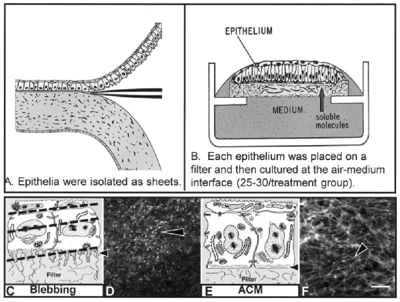
A,B: Schematic drawings of corneal epithelia isolated from the underlying stroma (A) and cultured at the air–medium interface (B). Soluble stimulating and inhibiting substances can be added to the medium for specific culture times. C: Epithelia isolated without the basal lamina extended basal cellular processes termed blebs as shown in the schematic drawing (arrowhead). D: The blebs contain F-actin (labeled with fluorescein isothiocyanate-phalloidin) that appears as punctate spots in a single confocal optical section taken through the basal area illustrated in the schematic drawing (C, lower dashed line). E: Tissue cultured with soluble extracellular matrix molecules reorganize the basal actin into an actin cortical mat (ACM) illustrated in the schematic drawing (arrowhead). F: The bundles of F-actin (phalloidin labeled) viewed in a single optical section from epithelia stimulated with collagen for 2 hr appear to align from cell to cell, across the field. Scale bar = 20 μm in F (applies to D,F).