Figure 5.
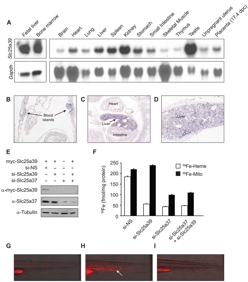
Mouse Slc25a39 is expressed in hematopoietic tissues and is important for heme biosynthesis. (A) Mouse tissue northern blot analysis of Slc25a39 expression. (B) Mouse Slc25a39 transcripts are localized to blood islands of the yolk sac at early somite stages (E8.5, arrows). (C) and (D) Slc25a39 transcripts accumulate most abundantly in the liver where expression is heterogeneous (E12.5). (E) and (F) MEL cells were differentiated for two days in media containing 1.5% DMSO prior to (E) transient transfection with myc-tagged Slc25a39 or (F) silencing of Slc25a39 (si-Slc25a39) and/or Slc25a37 (si-Slc25a37) using siRNA oligos. Assays were performed after two additional days of differentiation. si-NS indicates silencing using non-specific control oligos. (E) Representative western blot using anti-myc (α-myc-Slc25a39) and anti-Slc25a37 antibodies. Equal loading was verified by anti-tubulin. (F) MEL cells were metabolically labeled with 59Fe conjugated to transferrin, and total mitochondrial iron (59Fe-Mito) and iron in heme (59Fe-Heme) were determined. Results shown are from two independent experiments assayed in duplicate; error bars denote standard deviation. (G) (H) and (I) Representative photos of (G) an uninjected, wild-type control zebrafish embryo, (H) a zebrafish embryo injected with a ferrochelatase-specific morpholino, or (H) with a slc25a39-specific morpholino (I). Arrow indicates porphyric red blood cells in circulation.

