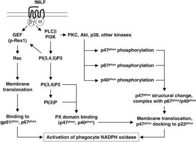
Fig. 6.
The fMLF-induced signaling events leading to the activation of phagocyte NADPH oxidase (Nox2). Depicted schematically are major signaling pathways activated by fMLF that results in NADPH oxidase activation in neutrophils. Upon activation of the Gαi proteins, the released Gβγ subunits trigger PI3K activation, resulting in the production PIP3 and its degradation products. The Gβγ and PIP3-mediated p-Rex1 activation is key to the conversion of GDP-bound Rac small GTPase to GTP-bound Rac, which translocates to membrane and associates with gp91phox.Gβγ is also responsible for PLCβ2 activation, leading to the production of the second mesengers diacyl glycerol and 1,4,5-inositol trisphosphate (IP3), which stimulate PKC activation. Isoforms of PKC (PKCδ, PKCξ, PKCβII, and PKCα), MAPK (p38, ERK), and Akt are known to catalyze the phosphorylation of p47phox in fMLF-stimulated neutrophils, prompting membrane translocation of the cytosolic factors. The PX domain in p47phox also facilitates its membrane association. Assembly of a membrane complex of NADPH oxidase is key to its conversion of molecular oxygen to superoxide. Omitted in this drawing is the phospholipase D (PLD) activation pathway, which is reported to contribute to fMLF-induced superoxide generation.

