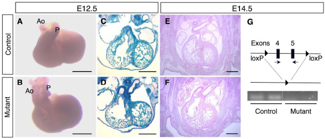
Figure 2. Outflow tract defects in BMPRIIflox/−;Mox2-Cre mice at E12.5–14.5.
(A, B) Photographs of embryonic hearts from control (A) and mutant (B) mice at E12.5. Ao: aorta, P: pulmonary artery. (C, D) Staining for β-galactosidase activity was performed to detect gene recombination on the frozen sections of the hearts from control (C) and mutant (D) mice at E12.5. (E, F) Hematoxylin and eosin staining of the embryonic hearts from control (E) and mutant (F) mice at E14.5. DORV was observed in the mutant hearts at E12.5 (D) and 14.5 (F). (G) Schematic diagram of the “floxed” allele and the deleted allele with loxP sites (filled triangles). BMPRII gene expression was detected by RT-PCR using a primer set for exons 4 and 5 (arrows). Ethidium bromide staining of PCR products for the BMPRII gene expression was shown for control and mutant (n=2 for each). Scale bars: 0.5 mm (A–F).

