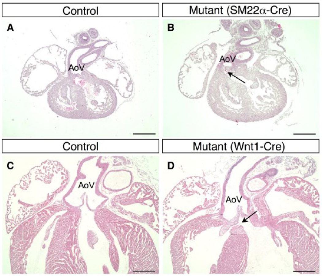
Figure 7. Overriding aorta in BMPRIIflox/−;SM22α-Cre and BMPRIIflox/−;Wnt1-Cre mice.
(A–D) Hematoxylin and eosin staining is shown for a control mouse (A) and a BMPRIIflox/−;SM22α-Cre mouse (B) at E14.5, and a control mouse (C) and a BMPRIIflox/−;Wnt1-Cre mouse (D) at P3. The OFT from the left ventricle was severely deviated to the right side of the heart in the BMPRIIflox/−;SM22α-Cre mouse. The aortic valve is located over the right ventricle (B, arrow) in this long axis cross section, whereas the aortic valve in a control mouse is located centrally (A). An overriding aorta with a small VSD (D, arrow) is observed in BMPRIIflox/−;Wnt1-Cre mice, whereas the normal position of the aorta is shown in control mice at P3 (C). AoV: aortic valve. Scale bars: 0.5 mm (A–D).

