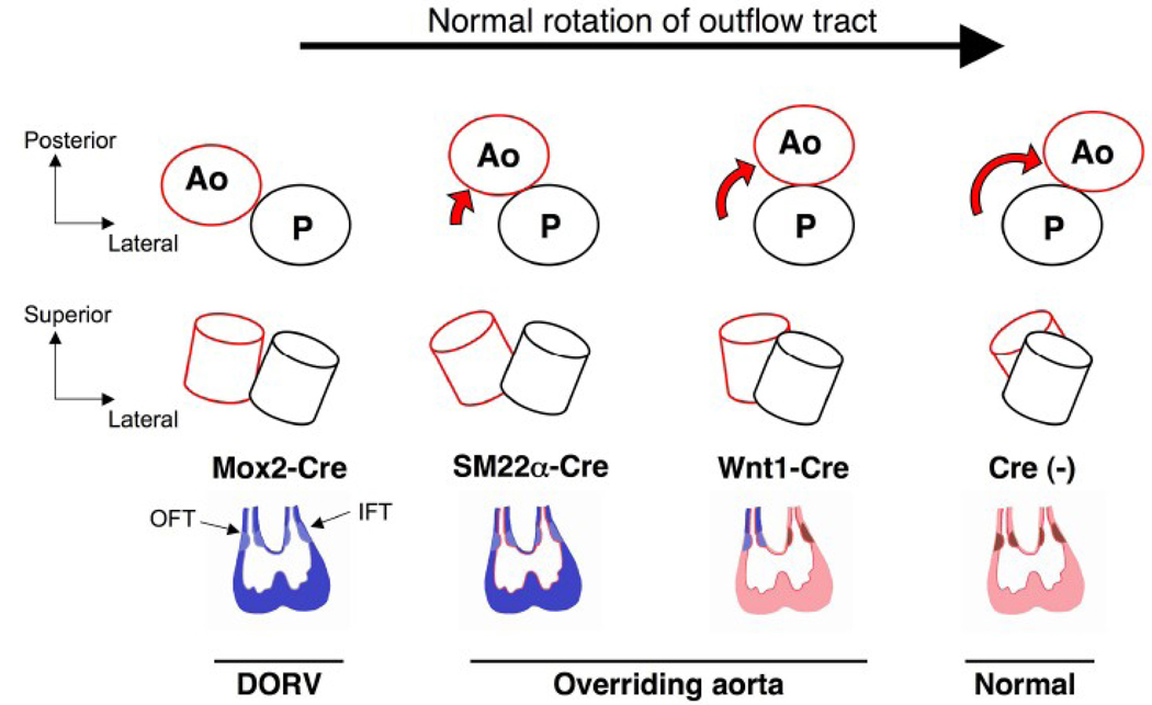
Figure 8. BMPRII deficiency interrupts normal rotation of the OFT in a tissue-specific manner.
The normal rotation of the aorta (red) and the pulmonary artery (black) during cardiac development is shown from a cranial view (upper panel) and a lateral view (middle panel). In this illustration of embryonic hearts (looping stage), BMPRII-deficient cardiac tissues are colored in blue for each Cre transgene (lower panel). Normal tissues (Cre-negative) are shown in pink (myocardium), red (endocardium), and brown (OFT and AV cushions). The rotational process is interrupted at different stages for each Cre transgenic strain as shown in the diagrams. Note that neural crest-derived cells expressing Wnt1-Cre partially contribute to the formation of the OFT cushion. Varying degrees of malrotation are observed with BMPRIIflox/− mice with the Mox2-Cre, SM22α-Cre, and Wnt1-Cre transgenes. The severity of aortic malpositioning likely correlates with the number of BMPRII-deficient mesenchymal cells in the OFT cushion and/or the timing of BMPRII deletion. Ao: aorta, P: pulmonary artery, IFT: inflow tract, OFT: outflow tract.

