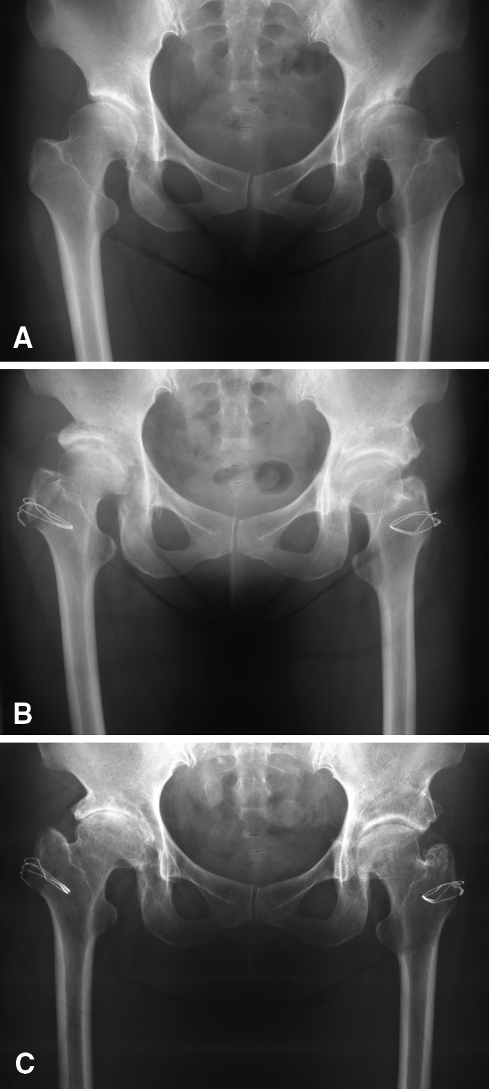
Fig. 1A–C.

(A) An AP radiograph of the pelvis shows marked bilateral hip dysplasia of a symptomatic 53-year-old woman who had previous acetabular osteotomies. (B) A postoperative AP radiograph of the patient’s right hip at 1 month and of the left hip at 1 year shows marked improvement in femoral head coverage. The patient had marked improvement in function. (C) Fifteen years after surgery, the patient’s postoperative AP radiograph shows substantial deterioration of the joint space of the right hip but preservation of the joint space of the left hip.
