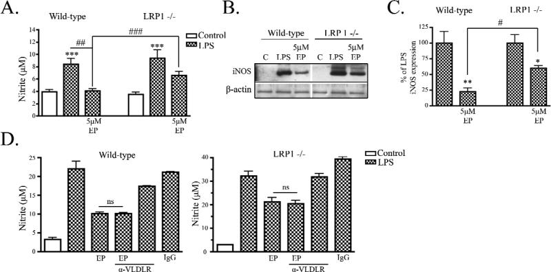
Figure 3. ApoE peptide attenuated LPS-induced NO production in LRP1 expressing microglia.
A) Wild-type and LRP1 −/− microglia were treated with 100 ng/mL LPS and nitrite was measured in the conditioned media at 24 h. LPS induced nitrite accumulation in both cell types and EP showed attenuation of LPS-induced nitrite production only in wild-type cultures (mean ± SEM; *** P < 0.001 compared to corresponding control cultures; ## P < 0.01, ### P < 0.001 compared with indicated cultures; n = 6 for each of 4 independent experiments). B) Cell lysates from wild-type and LRP1 −/− cultures were analyzed by Western blotting for iNOS and b-actin. Representative blots show that EP and LPS treatment reduced iNOS expression compared to LPS stimulation alone in wild-type stimulated cultures but not LRP1 −/− cultures. C) Western blot data were quantified as percent of control (mean ± SEM; * P < 0.05, ** P < 0.01 compared with LPS stimulated cultures; # < 0.05 compared with indicated cultures; n = 6 for each of 4 independent experiments). D) Wild-type (left panel) and LRP1 −/− (right panel) cultures were treated LPS and nitrite was measured in the conditioned media at 24 h. LPS induced nitrite accumulation in wild-type and LRP1 −/− cultures. EP and LPS treatment attenuated LPS-induced nitrite accumulation significantly in wild-type cultures and treatment with 500 nM VLDLR blocking antibody, EP and LPS did not prevent the effects of EP. Similarly in LRP1 −/− microglia, nitrite accumulation in cultures treated with VLDLR antibody cocktail did not significantly differ from cultures treated with EP and LPS (mean ± SEM; ns means not significant; n = 4).

