
Figure 2.
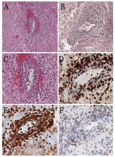
Histopathologic evaluation of vasculitic tissue from the brain. H & E stained sections of autopsy material from the brain of Patient No. 1 demonstrate perivascular lymphohistiocytic infiltrates (A). The lymphocytes are mostly small and round, with benign appearance; scattered cells are medium to large sized (C). Reticulin staining highlights the mononuclear infiltrate within vessel walls and adventitia in a “tree ring” fashion (B). The inflammatory infiltrate is composed of predominantly CD8+ (D), and CD3+ (E) T-lymphocytes. The T-cells show aberrantly decreased or loss of CD5 staining (F).