
Figure 5. Distribution of the total (A), type I (B) and type II (C) IL-18Rα mRNAs in the cerebral cortex, in the hypothalamus and in the cerebellum of C57BL6/J mice.
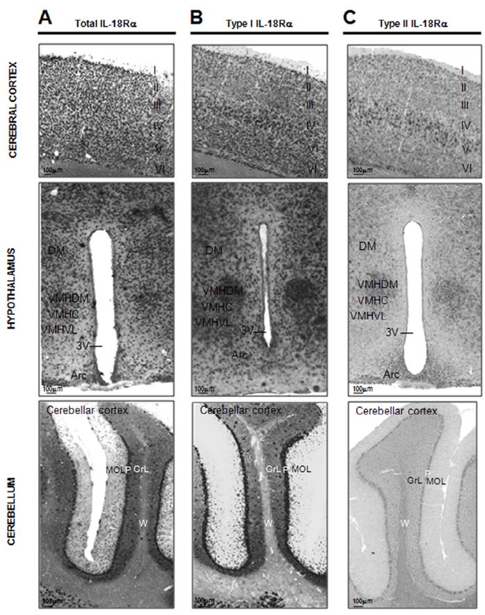
Upper panel shows distribution of the total (A), type I (B) and II (C) IL-18Rα mRNAs in the somatosensory cortex of C57BL6/J mice demonstrated with a DIG-labeled antisense cRNA probes- I: molecular layer, II: external granular layer, III external pyramidal layer, IV: internal granular layer, V: internal pyramidal layer, VI: multiform layer; medium panel shows distribution of the total (A), type I (B) and II (C) IL-18Rα mRNAs in the hypothalamus of C57BL6/J mice demonstrated with a DIG-labeled antisense cRNA probes. DM: dorsomedial hypothalamic nucleus, VMHC: ventromedial hypothalamic nucleus central part, VMHDM: ventromedial hypothalamic nucleus dorsomedial part, VMHVL: ventromedial hypothalamic nucleus ventrolateral part, 3V: third ventricle, Arc: arcuate hypothalamic nucleus; lower panel shows distribution of the total (A), type I (B) and II (C) IL-18Rα mRNAs in the cerebellar cortex of the cerebellum of C57BL6/J mice demonstrated with a DIG-labeled antisense cRNA probes- MOL: molecular layer, P: Purkinje cell layer, GrL: granular layer, W: white matter, of the cerebellar cortex.