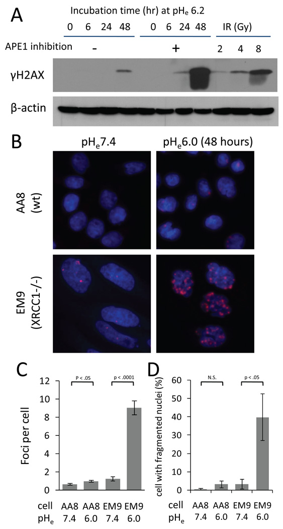
Fig. 5.
(A) The time-course of phosphorylation of histone H2AX by Western blotting in HCT116 cells treated with 0µM or 600µM CRT0044876 at pHe6.2. For comparison, the extent of H2AX phosphorylation at 30 min following ionizing radiation (IR) is also shown. (B) Immunocytochemical analyses for γH2AX foci formation (red) (×640). Nuclei were counterstained with Hoechst33342 (blue). A 48-hour incubation at pHe6.0 induced greater γH2AX foci formation in CHO EM9 cells compared to CHO AA8 cells. (C) Quantitative analysis of γH2AX foci formation in >500 nuclei from 5 independent experiments (mean±sem). (D) The number of cells with micronuclei was counted in the same experiments shown in the panel B and C (mean±sem). Significance of difference between the two groups was tested using t-test.

