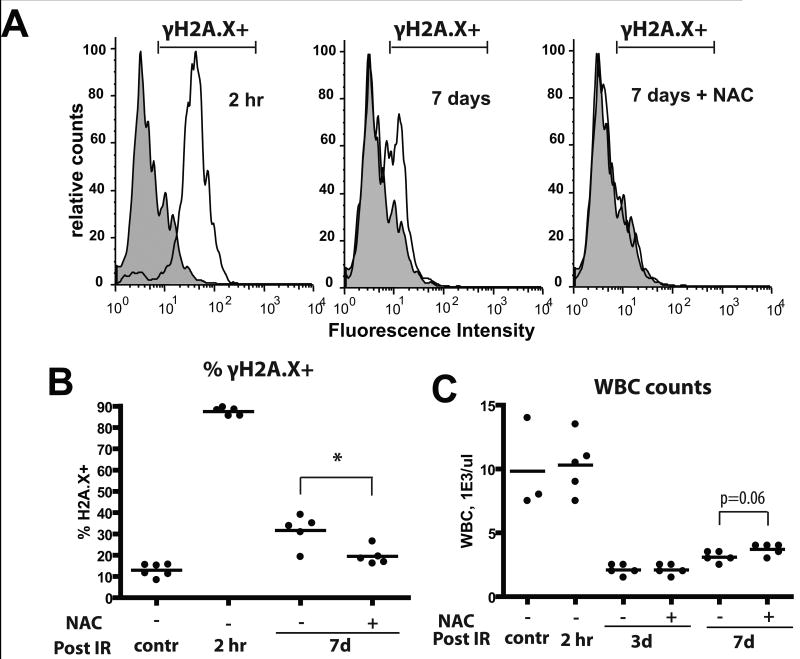
Figure 3. X-irradiation of mice results in persistent increases in DNA damage in BM cells.
Mice were irradiated at 5 Gy and transferred into cages with water +/- 40μM NAC. At the indicated time-points, mice were sacrificed and BM harvested. A) BM cells were permeabilized and stained with a fluorescent-linked antibody to γH2AX. Representative flow profiles are shown (gray overlay plot: analysis of untreated/control BM). B) Percentages of γH2AX+ cells at the indicated time-points are plotted (5-6 mice/group). C) Leukocyte counts from peripheral blood were determined using automated CBC analysis.

