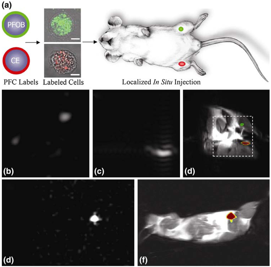
FIGURE 5.
Localization of labeled cells after in situ injection. (a) To determine the utility for cell tracking stem/progenitor cells labeled with either PFOB (green) or CE ( red), nanoparticles were locally injected into mouse thigh skeletal muscle. (b–d) At 11.7 T, spectral discrimination permits imaging the fluorine signal attributable to ~1 × 10⁶ PFOB-loaded (b) or CE-loaded cells (c) individually, which when overlaid onto a conventional 1H image of the site (d) reveals PFOB and CE labeled cells localized to the left and right leg, respectively (dashed line indicates 3 × 3 cm2 field of view for 19F images). (e, f) Similarly, at 1.5 T, 19F image of ~4 × 10⁶ CE-loaded cells (e) locates to the mouse thigh in a 1H image of the mouse cross section (f). The absence of background signal in 19F images (b, c, e) enables unambiguous localization of perfluorocarbon-containing cells at both 11.7 T and 1.5 T. (Reprinted with permission from Partlow et al.40)

